
Endometrium facts for kids
The endometrium is the special lining inside a girl's or woman's uterus. Think of the uterus as a pear-shaped organ where a baby can grow. The endometrium is the soft, inner layer of this organ. It's very important because it gets ready each month to welcome a fertilized egg. If an egg isn't fertilized, this lining is shed during a girl's monthly period.
What is the Endometrium?
The endometrium is like a cozy bed that forms inside the uterus. It's made of special cells and blood vessels. Its main job is to prepare for a possible pregnancy. Every month, this lining grows thicker and richer with blood. This makes it a perfect place for a tiny fertilized egg to attach and start growing into a baby.
Monthly Changes
The endometrium changes a lot during a girl's menstrual cycle. This cycle is controlled by special chemicals called hormones.
- Building Up: At the start of the cycle, the endometrium begins to grow and thicken. It gets ready for a fertilized egg.
- Waiting: If a fertilized egg arrives, it can implant itself in this thick lining.
- Shedding: If no fertilized egg arrives, the body doesn't need the thick lining anymore. It breaks down and is shed from the body. This shedding is what we call a menstrual period. It's a normal and healthy part of a girl's body.
Why is it Important?
The endometrium is vital for reproduction. It plays a key role in:
- Pregnancy: Providing a safe and nourishing place for a fertilized egg to grow.
- Menstruation: Allowing the body to reset each month if pregnancy doesn't happen. This process keeps the reproductive system healthy and ready for the future.
Images for kids
-
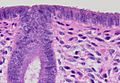
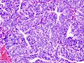
Histology of the most superficial layer of the endometrium, consisting of a simple columnar epithelium. H&E stain
-
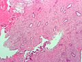
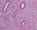
Low magnification micrograph of decidualized endometrium. H&E stain
-
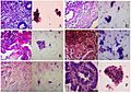
Histopathologic and cytopathologic images. (A) proliferative endometrium (Left: HE × 400) and proliferative endometrial cells (Right: HE × 100) (B) secretory endometrium (Left: HE × 10) and secretory endometrial cells (Right: HE × 10) (C) atrophic endometrium (Left: HE × 10) and atrophic endometrial cells (Right: HE × 10) (D) mixed endometrium (Left: HE × 10) and mixed endometrial cells (Right: HE × 10) (E): endometrial atypical hyperplasia (Left: HE × 10) and endometrial atypical cells (Right: HE × 200) (F) endometrial carcinoma (Left: HE × 400) and endometrial cancer cells (Right: HE × 400).
-
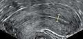
Triple-line endometrium measuring 7mm.
-
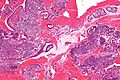
Endometrioid adenocarcinoma from biopsy. H&E stain.
-
Micrograph of decidualized endometrium due to exogenous progesterone. H&E stain.
-
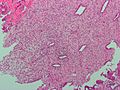
Micrograph of decidualized endometrium due to exogenous progesterone. H&E stain.
-
Micrograph showing endometrial stromal condensation, a finding seen in menses.


.jpg)


See also
 In Spanish: Endometrio para niños
