
Skin condition facts for kids
Quick facts for kids Skin condition |
|
|---|---|
| Synonyms | Cutaneous condition |
 |
|
| 3D medical illustration showing major layers of skin | |
| Causes | Germs (like bacteria and viruses), tiny bugs, injuries, serious illnesses, allergies, sun, heat or cold, dryness, too much moisture, problems with blood flow, ingrown hairs or nails, inherited traits, and other health issues. |
A skin condition is any health issue that affects your integumentary system. This system is like your body's protective suit. It includes your skin, nails, and important glands. Its main job is to act as a shield against the outside world.
There are many different kinds of skin conditions. Some are very common, while others are rare. Doctors often group these conditions by where they appear on the body, what they look like, or what caused them. To figure out what a skin condition is, doctors look closely at the skin. They check its location, how it feels (like if it's itchy or painful), how long it's been there, and its shape and color. Sometimes, they might take a tiny sample of skin to look at under a microscope. This helps them understand the condition better.
Contents
Your Amazing Skin: A Protective Shield
Your skin is the largest organ in your body! It weighs about 4 kilograms (9 pounds) and covers an area of about 2 square meters (21 square feet). It's made up of three main layers that work together to protect you.
What is Your Skin Made Of?
Your skin has three distinct layers: the epidermis, the dermis, and the subcutaneous tissue. These layers help your body in many ways, from keeping out germs to controlling your temperature.
The Epidermis: Your Outer Layer
The epidermis is the very top layer of your skin. It's like a strong, thin wall. This layer has several parts, but the most important cells here are keratinocytes. These cells make a tough protein called keratin, which helps protect your skin. New skin cells are always forming at the bottom of the epidermis. They slowly move up to the surface, where old cells are shed. This process takes about four weeks. The epidermis also contains melanocytes, which are cells that make melanin. Melanin is the pigment that gives your skin, hair, and eyes their color. It also helps protect your skin from the sun.
The Dermis: The Middle Layer
The dermis is the layer right below the epidermis. It's thicker and contains many important things. Here, you'll find strong fibers like collagen and elastic fibers. These make your skin strong and stretchy. The dermis also has hair follicles (where hair grows), sebaceous glands (which make oil to keep your skin soft), and sweat glands (which help cool you down). There are also lots of tiny blood vessels in the dermis. They bring nutrients to your skin, help control your body temperature, and assist in healing wounds.
The Subcutaneous Tissue: The Deepest Layer
The subcutaneous tissue is the layer of fat and other tissues beneath the dermis. It's like a cushion for your body. This layer helps to insulate you, keeping you warm. It also absorbs shocks and bumps, protecting your internal organs. Plus, it stores energy for your body to use later. The main cells in this layer are adipocytes, which are fat cells.
What Are Skin Conditions?
Skin conditions are health issues that affect your skin, hair, or nails. They can range from common problems like acne to more serious issues. Some conditions are caused by infections from germs, while others might be unusual growths.
How Doctors Understand Skin Conditions
When a doctor examines your skin, they look for specific changes. These changes are often called "lesions." Doctors pay attention to what the lesions look like, how they are grouped, and where they are on your body. The first type of change that appears is called a "primary lesion." As a condition develops or is affected by scratching, these can turn into "secondary lesions." Understanding these changes helps doctors figure out the best way to help you.
Looking at Skin Changes: Lesions
Skin lesions are simply changes in the skin that look different from the surrounding area. They can be a sign of a skin condition.
Primary Lesions: The First Signs
These are the original changes that appear on your skin.
- Macule: A flat spot on the skin that has a different color. It's not raised or sunken. Think of a freckle!
- Patch: Like a macule, but larger. It's a big, flat area of changed skin color.
- Papule: A small, solid bump on the skin. It's usually less than 10 mm (about the size of a pencil eraser).
- Plaque: A larger, flat-topped raised area. It's like a broad papule.
- Nodule: A solid, round bump that is deeper in the skin than a papule.
- Tumor: A larger nodule, bigger than 10 mm. It's a general term for a growth.
- Vesicle: A small blister filled with clear fluid. It's usually less than 10 mm.
- Bulla: A large blister filled with clear fluid. It's bigger than a vesicle.
- Pustule: A small bump on the skin filled with pus (a mix of inflamed cells and fluid).
- Cyst: A sac-like pocket in the skin that contains fluid or other material.
- Wheal: A raised, itchy, red or pale bump that appears suddenly and usually disappears within a day or two. Think of a hives bump.
- Telangiectasia: Tiny, visible blood vessels that look like fine red lines on the skin.
- Burrow: A thin, wiggly line on the skin caused by tiny organisms digging tunnels.
Secondary Lesions: Changes Over Time
These lesions develop from primary lesions or are caused by scratching or healing.
- Scale: Flaky, dry, or oily bits of skin that come off the surface.
- Crust: Dried fluid (like blood or pus) on the skin's surface. Think of a scab.
- Lichenification: Skin that has become thick, tough, and leathery, often from constant scratching.
- Erosion: A shallow open sore where only the top layer of skin (epidermis) is missing.
- Excoriation: A scratch mark on the skin, often from scratching an itch.
- Ulcer: A deeper open sore where both the epidermis and part of the dermis are missing.
- Fissure: A narrow, deep crack in the skin.
- Induration: Skin that feels unusually thick and firm.
- Atrophy: Thinning of the skin, which can make it look wrinkled or sunken.
- Maceration: Skin that becomes soft and white from being wet for too long.
How Skin Changes Are Arranged
This describes how lesions are grouped together on your skin.
- Annular or circinate: Shaped like a ring.
- Arciform or arcuate: Shaped like an arc or half-moon.
- Linear: In a straight line.
- Reticular or reticulated: Shaped like a net or web.
- Stellate: Star-shaped.
- Targetoid: Looks like a bullseye target.
Where Skin Changes Appear
This describes the pattern of where lesions are located on your body.
- Generalized: Spread over a large area of the body.
- Symmetric: Appearing on both sides of the body in a similar way.
- Flexural: Found in skin folds, like the elbows or knees.
- Extensor: Found on the outer surfaces of joints, like the back of the elbows or knees.
- Intertriginous: In areas where skin rubs together, like under the arms.
- Palmoplantar: On the palms of the hands or soles of the feet.
- Photodistributed: Appearing in areas exposed to sunlight.
- Zosteriform or dermatomal: Following the path of a nerve, often on one side of the body.
A Little Bit of History
Did you know that the study of skin conditions has a long history? In 1572, a doctor named Geronimo Mercuriali from Italy wrote a book called De morbis cutaneis. This book is considered the very first scientific work dedicated to dermatology, which is the study of skin diseases.
Images for kids
-
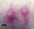
Chigger bites on human skin showing characteristic welts
-
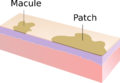
Macule and patch
-
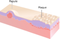
Papule and plaque
-
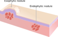
Nodules
-
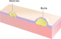
Vesicles and bulla
-
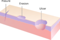
Fissures, erosions and ulcers
-
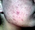
A pustule on the cheek
-
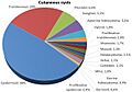
Relative incidence of skin cysts


