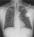
Chest x-ray facts for kids
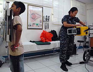
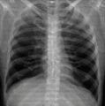
A chest X-ray (also called a CXR or chest radiograph) is a special picture of your chest. Doctors use it to look inside your body without needing to do surgery. It helps them check your chest, including your lungs, heart, and bones.
Chest X-rays are one of the most common types of medical images taken. They use a small amount of X-rays (a type of energy) to create pictures of your insides. This helps doctors find out what might be making you sick.
What Can a Chest X-ray Show?
Doctors use chest X-rays to find many different problems in the chest area. They can see your bones, lungs, heart, and the large blood vessels near your heart.
Some common conditions that a chest X-ray can help find include:
- Pneumonia: This is an infection in your lungs.
- Pneumothorax: This happens when air leaks into the space around your lung.
- Congestive heart failure: This is when your heart doesn't pump blood as well as it should.
- Broken bones: Like a broken rib.
- Hiatal hernia: This is when part of your stomach pushes up into your chest.
Doctors also use chest X-rays to check for lung diseases related to certain jobs. For example, miners who breathe in dust might get regular X-rays.
When doctors look at a chest X-ray, they check different parts. They look at your:
- Airways: The tubes that carry air to your lungs.
- Bones: Like your ribs and spine.
- Cardiac silhouette: The outline of your heart to see if it's too big.
- Diaphragm: The muscle under your lungs that helps you breathe.
- Edges: The very top and sides of your lungs.
- Fields: The main parts of your lungs to see if there's any fluid or damage.
What a Chest X-ray Might Not Show
Chest X-rays are helpful and generally safe. However, they don't always show every problem. Sometimes, a person can have a serious chest condition, but their X-ray looks completely normal.
For example, if someone is having a heart attack, their chest X-ray might look fine. In these cases, doctors might need to do other tests to find out what's wrong.
Images for kids
-
A chest X-ray with different parts of the ribs and other areas labeled.
-
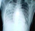
This chest X-ray shows a clear wedge-shaped area in the right lung, which is a sign of pneumonia.
-
A CT scan showing how chest structures look from different angles.
-
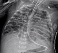
This chest X-ray shows increased cloudiness in both lungs, which can mean pneumonia.
 | Kyle Baker |
 | Joseph Yoakum |
 | Laura Wheeler Waring |
 | Henry Ossawa Tanner |


