
Electrophysiology facts for kids
Electrophysiology is the study of the electrical signals inside living things. It looks at how cells and tissues use electricity. Scientists measure tiny electrical signals, from a single tiny protein to a whole organ like your heart.
In neuroscience, electrophysiology helps us understand how neurons (brain cells) send electrical messages. These messages are often called action potentials.
Contents
How We Study Electricity in Living Things
To study these electrical signals, scientists use special tools called electrodes. Electrodes are like tiny sensors that can pick up electrical signals. They can be simple metal discs or needles. Some are even hollow glass tubes filled with a special liquid.
Scientists place these electrodes into different parts of living things. This can be:
- A living animal.
- A piece of excised tissue (tissue removed from an animal).
- Single cells grown in a lab.
- Artificially grown cells or tissues.
If an electrode is very small, a scientist can even put its tip inside a single cell. This lets them measure the electricity of that one cell.
What Electrophysiology Measures
Many specific electrophysiological readings have special names, depending on what part of the body they are studying:
- Electrocardiography (ECG or EKG) – measures the electrical activity of the heart.
- Electroencephalography (EEG) – measures electrical activity in the brain.
- Electrocorticography (ECoG) – measures electrical activity directly from the surface of the cerebral cortex (the outer part of the brain).
- Electromyography (EMG) – measures the electrical activity of muscles.
- Electrooculography (EOG) – measures the electrical activity of the eyes.
- Electroretinography (ERG) – measures the electrical activity of the retina (the light-sensing part of the eye).
- Electroantennography (EAG) – measures the electrical activity of smell receptors in insects like moths.
Images for kids
-
"Current Clamp" is a common technique in electrophysiology. This is a whole-cell current clamp recording of a neuron firing due to it being depolarized by current injection
-
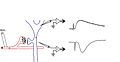
A schematic diagram showing a field potential recording from rat hippocampus. At the left is a schematic diagram of a presynaptic terminal and postsynaptic neuron. This is meant to represent a large population of synapses and neurons. When the synapse releases glutamate onto the postsynaptic cell, it opens ionotropic glutamate receptor channels. The net flow of current is inward, so a current sink is generated. A nearby electrode (#2) detects this as a negativity. An intracellular electrode placed inside the cell body (#1) records the change in membrane potential that the incoming current causes.
-
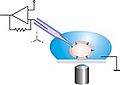
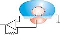
Schematic drawing of the classical patch clamp configuration. The patch pipette is moved to the cell using a micromanipulator under optical control. Relative movements between the pipette and the cell have to be avoided in order to keep the cell-pipette connection intact.
-
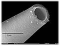
Scanning electron microscope image of a patch pipette.
-
In planar patch configuration, the cell is positioned by suction. Relative movements between cell and aperture can then be excluded after sealing. An antivibration table is not necessary.
-
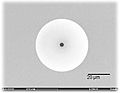
Scanning electron microscope image of a planar patch clamp chip. Both the pipette and the chip are made from borosilicate glass.


See also
 In Spanish: Electrofisiología para niños
