History of cell membrane theory facts for kids
The cell membrane is like a tiny, invisible fence that surrounds every cell. It controls what goes in and out, keeping the cell safe and working properly. Scientists first saw cells with microscopes in the 1600s. But it took almost 200 years to figure out what this amazing barrier was made of!
By the 1800s, everyone agreed that cells must have some kind of special boundary. Early ideas suggested it might be made of fat, also called lipids. Then, in 1925, experiments showed that this barrier was actually two layers of lipids, called a lipid bilayer. Over time, new tools helped confirm this idea.
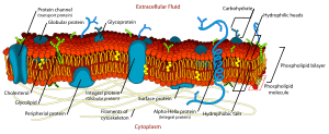
However, there was still a big question: what about proteins? How did they fit into the cell membrane? Eventually, scientists came up with the fluid mosaic model. This model suggests that proteins "float" like icebergs in a "sea" of fluid lipid layers. Even though it's a bit simplified, this model is still very important today for understanding cells.

Contents
Early Ideas About the Cell's Barrier
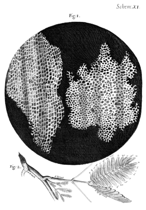
When microscopes were invented in the 1600s, scientists like Robert Hooke discovered that plants and animals are made of tiny cells. Plant cells had a clear outer wall, but animal cells didn't seem to have one. Still, scientists knew there had to be a barrier.
By the mid-1800s, scientists like Moritz Traube realized this outer layer had to be "semipermeable." This means it lets some things pass through, like water, but blocks others. Traube thought it was formed when the cell's inside stuff mixed with the fluid outside.
The correct idea that the cell membrane was made of lipids (fats) came from Georg Hermann Quincke in 1888. He noticed that cells in water are round, like oil drops. If a cell broke, it formed two smaller round cells, just like oil. He also saw that a thin film of oil acts like a semipermeable membrane. Quincke believed the cell membrane was a thin, fluid layer of fat, less than 100 nanometers thick.
More evidence came from studying anesthetics. Hans Horst Meyer and Ernest Overton found that chemicals used as anesthetics could dissolve in both water and oil. They thought this meant a molecule had to be able to dissolve in oil to get past the cell membrane. They called this their "lipoid theory of narcosis." They even suggested the membrane might be made of lecithin and cholesterol.
One problem with this early idea was that it didn't explain how cells could actively move certain things in or out. This mystery was solved much later. Scientists found special molecules called integral membrane proteins that act like tiny pumps, moving things across the membrane.
Discovering the Lipid Bilayer Structure
By the early 1900s, scientists knew the cell membrane was likely made of lipids. But they didn't know its exact structure. Two important experiments in 1924 helped figure this out.
A scientist named Fricke measured the electrical properties of red blood cells. He calculated that the cell membrane was about 3.3 nanometers thick. However, he thought it was just one layer of molecules. He actually measured only the middle part of the membrane, not the whole thing.
Then, Gorter and Grendel tried a different approach. They extracted lipids from red blood cells and spread them out in a single layer. They found that the area of this lipid layer was twice the surface area of the original cells. Even though their experiment had a few small errors, their main conclusion was correct: the cell membrane is a lipid bilayer, meaning it has two layers of lipids.
About ten years later, Davson and Danielli added to this idea. They suggested that the lipid bilayer was covered on both sides by a layer of globular proteins. They thought these proteins just stuck to the membrane. However, they were wrong about how the proteins worked and why the membrane acted as a barrier.
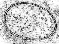
In the late 1950s, electron microscopy allowed scientists to see the membrane more directly. After staining cells, scientists saw two dark lines separated by a lighter area. At first, they thought the dark lines were a single protein layer. But J. David Robertson correctly realized that the dark lines were the ends of the two lipid layers, along with any attached proteins. He called this the "unit membrane," suggesting that all cell membranes, and even membranes inside organelles, had this same bilayer structure.
How the Membrane Theory Grew
Around the same time, the idea of a semipermeable membrane became important. This is a barrier that lets water pass through but blocks larger molecules. The word "osmosis" (the movement of water across a membrane) was first used in 1827.
In 1877, Wilhelm Pfeffer proposed the membrane theory of cell physiology. He said that cells were surrounded by a thin plasma membrane. He believed that the water and other substances inside the cell were like a weak solution.
Later, in 1889, Hamburger studied how red blood cells swelled when placed in different solutions. By measuring how fast they swelled, he could estimate how quickly substances entered the cells. He also found that about half of a red blood cell's volume was not water that could dissolve things.
Ernest Overton first suggested the idea of a lipid (oil) plasma membrane in 1899. But his idea didn't explain why water could pass through the membrane so easily. So, Nathansohn (1904) proposed the "mosaic theory." He thought the membrane wasn't just pure lipid, but a mix of lipid areas and areas with a semipermeable gel. Ruhland improved this idea, adding "pores" (tiny holes) for small molecules to pass through.
Since membranes are usually less permeable to negatively charged particles (anions), Leonor Michaelis thought that ions stuck to the walls of the pores. This would change how easily other ions could pass through. Michaelis also showed that there was an electrical difference across the membrane (called membrane potential) and linked it to how ions were spread out.
In 1939, Harvey and James Danielli suggested a lipid bilayer covered on each side with a layer of protein. This helped explain measurements of surface tension. In 1941, Boyle and Conway showed that the membrane of resting frog muscle let both potassium (K+) and chloride (Cl-) ions pass through, but not sodium (Na+). This meant that a single pore size could explain which ions passed through, without needing electrical charges in the pores.
The Idea of the Cell's "Pump"
With new tools like radioactive tracers, scientists found that cells are not completely impermeable to sodium (Na+). This was hard to explain with just a simple membrane barrier. So, the idea of a "sodium pump" was proposed. This pump would constantly remove sodium from cells as it entered.
This led to the concept that cells are always active, using energy to keep different amounts of ions inside and outside. In 1935, Karl Lohmann discovered ATP, which is the main energy source for cells. This discovery supported the idea of a metabolically-driven sodium pump.
The work of Hodgkin, Huxley, and Katz greatly supported the membrane pump idea. They developed mathematical models that correctly described the electrical signals across cell membranes.
Today, we know the plasma membrane is a fluid lipid bilayer with many different proteins embedded within it. We understand its structure in great detail, including 3D models of hundreds of these proteins. These discoveries showed how important the membrane theory is in understanding how cells work.
The Fluid Mosaic Model
Around the same time, scientists created the first "model membrane" by "painting" a lipid solution across a small opening. Mueller and Rudin found that this artificial membrane was fluid, had high electrical resistance, and could even repair itself if punctured. This type of model became known as a "BLM," which could mean "black lipid membrane" or "bimolecular lipid membrane."
In 1970, Frye and Edidin showed that this fluidity also happens on real cell surfaces. They mixed two cells with different colored fluorescent tags on their membranes. They watched as the colors mixed, proving that the membrane components could move around.
These results were key to the "fluid mosaic" model of the cell membrane. This model was developed by Singer and Nicolson in 1972. It says that biological membranes are mostly a lipid bilayer. Proteins are either partly or fully stuck into this bilayer. These proteins are thought to float freely in the liquid-like bilayer.
This wasn't the first time someone suggested a mixed membrane structure. Nathansohn had proposed a "mosaic" of water-permeable and impermeable areas in 1904. But the fluid mosaic model was the first to correctly combine the ideas of fluidity, membrane channels, and how proteins connect to the bilayer into one complete theory.
Modern Research
Even though the fluid mosaic model is great, later research found some things that needed more detail. For example, channel proteins were once thought to have a continuous water channel through their center. We now know this is usually not true, except for things like nuclear pore complexes.
Also, things don't always spread freely across the entire cell surface. Their movement is often limited to small areas. This is because of things like the cytoskeleton (the cell's internal "skeleton"), how lipids separate into different areas, and groups of proteins sticking together.
Today's studies also suggest that much less of the plasma membrane is "bare" lipid than once thought. In fact, a lot of the cell surface might be connected to proteins. Despite these small changes, the fluid mosaic model is still a very popular and important idea for understanding the structure of biological membranes.
Obsolete theories
Over time, some older ideas about cell membranes were replaced by the modern fluid-mosaic model. This model says that a lipid bilayer separates the inside from the outside of cells. It also explains that special channels, pumps, and transporters in the membrane control what goes in and out.
One older idea came from Gilbert Ling. He believed that the cell's internal jelly-like substance, called protoplasm, was responsible for how cells behaved, including their electrical properties and what could pass through them. The modern view, however, gives most of this credit to the plasma membrane.
As the lipid bilayer membrane theory gained support, Ling's alternative idea, which downplayed the membrane's importance, was largely rejected. Scientists like Procter & Wilson (1916) and Loeb (1920) showed that gels, which don't have a semipermeable membrane, could swell in water and even create an electrical difference. This suggested that some properties thought to belong to the membrane might also be found in the cell's internal material.
In the 1930s, some scientists criticized the membrane theory. For example, some cells could swell and increase their surface area by a huge amount. A lipid layer couldn't stretch that much without breaking. These criticisms encouraged more studies on protoplasm as the main thing controlling what enters and leaves the cell. Fischer and Suer (1938) suggested that water in the protoplasm was not free but combined with other chemicals. They showed that living tissues swelled similarly to gelatin gels. Dimitri Nasonov (1944) also thought proteins were key to many cell properties, including electrical ones.
By the 1940s, these "bulk phase theories" (ideas focusing on the cell's internal material) were not as well developed as the membrane theories and were mostly rejected. In 1941, Brooks & Brooks published a book that clearly rejected the bulk phase theories.
See also
 In Spanish: Historia de la teoría de la membrana celular para niños
 | Jewel Prestage |
 | Ella Baker |
 | Fannie Lou Hamer |


