Ophthalmology facts for kids

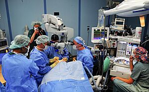
Ophthalmologists perform cataract surgery using an operating microscope
|
|
| System | Eye and visual system |
|---|---|
| Significant diseases | Cataract, retinal disease (including diabetic retinopathy and other types of retinopathy), glaucoma, corneal disease, Category:Disorders of eyelid, lacrimal system and orbit, uveitis, strabismus and disorders of the ocular muscles, ocular neoplasms (malignancies, or cancers, and benign eye tumors), neuro-ophthalmologic disorders (including disorders of the optic nerve) |
| Significant tests | Ophthalmoscopy, visual field test, optical coherence tomography |
| Specialist | Ophthalmologist |
| Occupation | |
|---|---|
| Names | Physician Surgeon |
|
Occupation type
|
Specialty |
|
Activity sectors
|
Medicine, surgery |
| Description | |
|
Education required
|
Doctor of Medicine (MD), Doctor of Osteopathic Medicine (DO), Bachelor of Medicine, Bachelor of Surgery (MBBS), Bachelor of Medicine, Bachelor of Surgery (MBChB) |
|
Fields of
employment |
Hospitals, Clinics |
Ophthalmology (pronounced OFF-thal-MOL-uh-jee) is a special field in medicine. It focuses on finding and treating problems with your eyes. It involves both medical treatments and surgery. Long ago, this field was sometimes called "oculism."
An ophthalmologist is a doctor who specializes in eye care. They go through extra training after medical school. This training teaches them how to treat eye diseases and perform eye surgeries. Many ophthalmologists also do research to learn more about eye conditions. This helps them find new ways to help people see better.
Contents
Common Eye Problems Ophthalmologists Treat
Ophthalmologists help people with many different eye conditions. Here are some common ones:
- Cataract: When the clear lens of the eye becomes cloudy.
- Excessive tearing: When your eyes water too much because tear ducts are blocked.
- Bulging eyes: When one or both eyes stick out more than usual.
- Diabetic retinopathy: Eye damage that can happen to people with diabetes.
- Dry eye syndrome: When your eyes don't make enough tears or the tears aren't good quality.
- Glaucoma: A group of diseases that can damage the optic nerve, often due to high pressure inside the eye.
- Macular degeneration: A condition that blurs the sharp, central vision needed for reading and driving.
- Retinal detachment: When the retina (the light-sensitive layer at the back of the eye) pulls away from its normal position.
- Refractive errors: Problems like nearsightedness or farsightedness that make vision blurry.
- Strabismus: When the eyes don't line up correctly, sometimes called "crossed eyes."
- Uveitis: Inflammation inside the eye.
- Eye trauma: Injuries to the eye from accidents.
How Eye Doctors Check Your Eyes
To find out what's going on with your eyes, ophthalmologists do several tests. These tests help them understand your vision and eye health.
Basic Eye Exam Methods
Here are some common ways eye doctors check your eyes:
- Visual acuity assessment: This checks how clearly you can see, often using an eye chart.
- Ocular tonometry: This measures the pressure inside your eye.
- Eye movement and alignment: This checks how well your eyes move together.
- Slit lamp examination: The doctor uses a special microscope to look closely at the front and inside of your eye.
- Dilated fundus examination: Eye drops are used to make your pupils wide. This lets the doctor see the back of your eye, including the retina and optic nerve.
- Refraction: This helps determine the right prescription for glasses or contact lenses.
Special Tools for Eye Checks
Doctors also use advanced tools for more detailed checks:
- Optical coherence tomography (OCT): This machine takes detailed pictures of the layers inside your eye. It helps doctors see problems like swelling or damage.
- Optical coherence tomography angiography (OCTA) and Fluorescein angiography: These tests take pictures of the blood vessels in your eye. They help find issues with blood flow.
- Electroretinography (ERG): This test measures the electrical signals from the light-sensing cells in your retina.
- Electrooculography (EOG): This measures electrical changes that happen when your eyes move. It helps check how well certain parts of your eye are working.
- Visual field testing: This checks your central and side vision. It can help find problems caused by conditions like glaucoma or even brain issues.
- Corneal topography: This creates a detailed map of the front surface of your eye (the cornea).
- Ultrasonography: Like an ultrasound for a baby, this uses sound waves to create images of the eye.
Eye Surgery
Eye surgery is when an ophthalmologist performs an operation on the eye or the areas around it. The eye is very delicate, so surgeons must be extremely careful. They choose the right surgery for each patient and make sure all safety steps are followed.
Special Areas in Eye Care
Ophthalmology has many subspecialties. These are like different branches that focus on specific diseases or parts of the eye.
- Anterior segment surgery: Operations on the front part of the eye.
- Cornea and outer eye diseases: Dealing with problems of the clear front part of the eye.
- Glaucoma: Focusing on the group of diseases that can damage the optic nerve.
- Neuro-ophthalmology: Eye problems related to the brain and nervous system.
- Ocular oncology: Treating eye cancers and tumors.
- Oculoplastics and orbit surgery: Surgery on the eyelids, tear ducts, and the area around the eye.
- Paediatric ophthalmology: Eye care for children, including treating strabismus (misaligned eyes).
- Refractive surgery: Surgeries to correct vision problems like nearsightedness, so people might not need glasses.
- Medical retina: Treating retinal problems without surgery.
- Uveitis: Treating inflammation inside the eye.
- Vitreo-retinal surgery: Surgical treatment for problems with the retina and the back part of the eye.
Sometimes, medical retina and vitreo-retinal surgery are combined into one specialty.
The History of Eye Care
People have been studying and treating eye problems for thousands of years.
Ancient Times
- In ancient Egypt, around 1550 BC, there were writings about eye diseases.
- Ancient Greek doctors like Aristotle studied animal eyes to learn about their structure. They realized the eye had layers and fluids.
- Later, Greek physicians like Rufus of Ephesus and Galen made more accurate drawings of the eye. They understood that the eye had different chambers and parts like the conjunctiva.
- The Greek philosopher Celsus described how to do cataract surgery using a method called "couching."
- In ancient India, the surgeon Sushruta wrote about 76 eye diseases and surgical tools around 600 BC. He was one of the first to describe cataract surgery.
Medieval Islamic Period
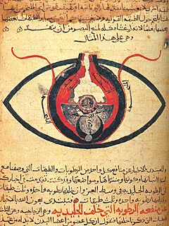
- Scientists in medieval Islamic countries combined theory with practical work. They made precise instruments for eye care.
- Doctors like Hunayn ibn Ishaq taught that the eye's lens was in the exact center.
- Ibn al-Nafis wrote a big textbook about eye theory and medicines.
- The word "retina" (for the back of the eye) came from this period.
Modern Eye Care
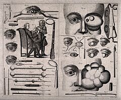
- In the 1600s and 1700s, scientists used new tools like microscopes to study the eye in more detail.
- Jacques Daviel performed the first planned cataract removal surgery in 1750.
- Georg Joseph Beer from Austria developed a new way to treat cataracts.
- Hermann von Helmholtz invented the ophthalmoscope in 1851. This amazing tool allowed doctors to look inside a living eye for the first time!
- Albrecht von Graefe was a very important ophthalmologist in the 1800s. He helped make ophthalmology a real medical specialty. He also developed new surgical techniques and treatments for glaucoma.
- Herman Snellen created the Snellen chart (the eye chart with letters) to test vision.
Famous Eye Doctors
Many brilliant doctors have helped advance eye care. Here are a few:
- Albrecht von Graefe (1828–1870) (Germany): A key founder of modern ophthalmology. He improved cataract surgery and glaucoma treatment.
- Allvar Gullstrand (1862–1930) (Sweden): Won a Nobel Prize for his research on how the eye focuses light.
- Hermann von Helmholtz (1821–1894) (Germany): Invented the ophthalmoscope, which lets doctors see inside the eye.
- Sir Harold Ridley (1906–2001) (United Kingdom): In 1949, he was one of the first to successfully put an artificial lens inside the eye after cataract surgery.
- Charles Schepens (1912–2006) (Belgium): Known as the "father of modern retinal surgery." He developed tools and techniques for treating retinal problems.
- Marshall M. Parks (1918–2005) (United States): Called the "father of pediatric ophthalmology" for his work with children's eye care.
- Charles Kelman (1930–2004) (United States): Developed a method called phacoemulsification for removing cataracts through a tiny cut.
Images for kids
See also
 In Spanish: Oftalmología para niños
- Book of the Ten Treatises of the Eye
- Chinese ophthalmology
- American Academy of Ophthalmology
- EyeWiki
- Eye disease
- Eye surgery
- Optometry
- Orthoptics
- Eye care professional


