Clot facts for kids
A blood clot is like a natural bandage your body makes. It's a thick, jelly-like clump of blood that forms to stop bleeding, especially when you get a cut. When you bleed, your blood changes into this clot at the injury site.
Sometimes, a blood clot is called a thrombus. The whole process of blood clotting is called coagulation.
If you get a cut on your body, you might start to bleed. To stop this, your body has an amazing system. First, your brain sends signals to your liver to make special chemicals. These chemicals travel to the injury and help the clot start to form. At the same time, your brain also helps slow down blood flow near the cut. It does this by making the blood vessels (like tiny tubes) in that area squeeze tighter. This means less blood is lost.
There's a limit to how fast a clot can form. If a cut is very deep and you bleed a lot, a clot might not be able to form quickly enough. This is why serious injuries need medical help.
How Blood Clots Form
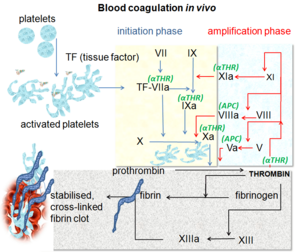
Blood clotting starts almost immediately after a blood vessel is hurt. This happens when the inner lining of the vessel, called the endothelium, gets damaged. When blood touches certain proteins, like tissue factor, it triggers changes in tiny blood cells called platelets and a protein in your blood called fibrinogen.
Your Platelets are like tiny repair workers. They quickly rush to the injury and form a plug, like a temporary seal. Then, other proteins in your blood, called coagulation factors or clotting factors, work together in a complex chain reaction. They create strong strands of a protein called fibrin. These fibrin strands act like glue, making the platelet plug much stronger and more stable.
This clotting system is very similar in all mammals, including humans. It involves both cells (platelets) and proteins (coagulation factors). Scientists have studied the human system the most, so we understand it best.
What is Fibrin?
Fibrin is a white, tough protein that doesn't dissolve in blood. It's made when another substance called thrombin acts on fibrinogen during clotting. Fibrin forms a strong net-like structure. This net then traps red blood cells and platelets, making the clot solid and helping to stop the bleeding.
Images for kids
-
Animation of the formation of an occlusive thrombus in a vein. A few platelets attach themselves to the valve lips, constricting the opening and causing more platelets and red blood cells to aggregate and coagulate. Coagulation of unmoving blood on both sides of the blockage may propagate a clot in both directions.
-
Micrograph showing a thrombus (center of image) within a blood vessel of the placenta. H&E stain.
-
Composition of a fresh thrombus at microscopy, showing nuclear debris in a background of fibrin and red blood cells.
 | Jessica Watkins |
 | Robert Henry Lawrence Jr. |
 | Mae Jemison |
 | Sian Proctor |
 | Guion Bluford |


