
Ventricular system facts for kids
| Brain: Ventricular system | ||
|---|---|---|
Quick facts for kids
 |
||
| The ventricular system helps make and move cerebrospinal fluid. | ||
 |
||
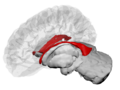
| A spinning 3D view of the four ventricles and their connections. From top to bottom: Blue - Lateral ventricles Cyan - Interventricular foramina (Monro) Yellow - Third ventricle Red - Cerebral aqueduct (Sylvius) Purple - fourth ventricle Green - connected to the central canal (Openings to subarachnoid space are not shown) |
||
| Latin | Ventriculi cerebri | |
The ventricular system is a group of four connected spaces inside your brain. These spaces are called cerebral ventricles. Inside each ventricle, there's a special area called the choroid plexus. This area makes a fluid called cerebrospinal fluid (CSF). This fluid moves around your brain and spinal cord. The ventricular system is connected to the central canal of the spinal cord. This allows the CSF to flow all around.
All parts of the ventricular system and the central canal are covered with a special lining. This lining is called ependyma. It's a type of epithelium with strong connections. These connections form a barrier that protects your brain.
Contents
How the Ventricular System Is Built

Your brain has four main ventricles:
- The lateral ventricles: There are two of these, one in each half of your brain.
- The third ventricle: This one is in the middle of your brain.
- The fourth ventricle: This is located at the back of your brain.
There are also several small openings called foramina. These act like channels that connect the ventricles. For example, the interventricular foramina connect the lateral ventricles to the third ventricle. This is how the cerebrospinal fluid moves between them.
| Name | Connects From | Connects To |
| interventricular foramina (Monro) | lateral ventricles | third ventricle |
| cerebral aqueduct (Sylvius) | third ventricle | fourth ventricle |
| median aperture (Magendie) | fourth ventricle | subarachnoid space (a space around the brain) |
| right and left lateral aperture (Luschka) | fourth ventricle | subarachnoid space |
The Ventricles Up Close

The four spaces in your brain are called ventricles. The two biggest ones are the lateral ventricles. They are found in the main part of your brain, the cerebrum. The third ventricle is in the center of your brain. It sits between two parts called the thalamus. The fourth ventricle is at the back of your brain. It is near the pons and medulla oblongata. All these ventricles work to make and move cerebrospinal fluid.
How the Ventricles Grow
The parts of the ventricular system start to form very early. They come from a tube-like structure called the neural canal. This canal is in the center of the neural tube, which later becomes your brain and spinal cord.
As your brain develops, this neural canal expands in certain areas. For example, the part that will become the brainstem expands to form the fourth ventricle. Other parts of the canal stay narrow, like the cerebral aqueduct. This aqueduct connects the third and fourth ventricles. The fourth ventricle then narrows down to become the central canal of your spinal cord.
Around the third week of development, a human embryo is like a flat disc. The top layer of cells is called the ectoderm. In the middle of this layer, a structure called the notochord forms. As the embryo grows, the brain starts to develop. By the fourth week, three main swellings appear around the neural canal. These swellings are called brain vesicles. They will become different parts of your central nervous system. As these parts grow, the inner neural canal becomes the "primitive" ventricles. These are the beginnings of your ventricular system.
What the Ventricular System Does
How Cerebrospinal Fluid Flows


The ventricles are filled with cerebrospinal fluid (CSF). This fluid surrounds and protects your brain and spinal cord. CSF is made by special cells in the choroid plexus. This plexus is found in most parts of the ventricular system.
CSF flows from the lateral ventricles. It then goes through the interventricular foramina into the third ventricle. From there, it moves into the fourth ventricle through the cerebral aqueduct. From the fourth ventricle, CSF can go into the central canal of the spinal cord. It can also flow into spaces around the brain called the subarachnoid cisterns. This happens through three small openings: one central opening and two side openings.
Most CSF is made by the choroid plexus. It then moves through the ventricles and spaces around the brain. Finally, it is absorbed back into your blood. This happens through tiny structures called arachnoid granulations. These structures are like small filters. The CSF then enters the venous sinuses and eventually the jugular vein. CSF can also flow down the spinal cord to the lower back. This is where doctors sometimes take samples of CSF using a procedure called a lumbar puncture.
The cerebral aqueduct and the foramina are quite small. This means they can sometimes get blocked.
Protecting Your Brain
Your brain and spinal cord are covered by three protective layers called the meninges. These layers are the tough dura mater, the arachnoid mater, and the pia mater. The cerebrospinal fluid (CSF) inside your skull and spine adds even more protection. It also helps your brain float. CSF is found in the subarachnoid space, which is between the pia mater and the arachnoid mater.
The CSF made in the ventricular system is also important for keeping your brain chemically stable. It provides nutrients that your brain needs. CSF helps protect your brain from bumps and jolts to your head. It also gives your brain buoyancy and support against gravity. Because your brain and CSF have similar densities, your brain floats. This allows your brain to grow without resting heavily on the bottom of your skull. If it did, it could damage brain tissue.
Health Issues Related to the Ventricular System
Because the cerebral aqueduct and other openings are narrow, they can sometimes get blocked. For example, blood from a stroke can block them. When CSF is constantly made but can't flow out, pressure builds up in the ventricles. This can lead to a condition called hydrocephalus. People sometimes call this "water on the brain." It is a very serious condition.
Doctors can perform a surgery called an endoscopic third ventriculostomy. In this surgery, a small opening is made in the third ventricle. This allows the cerebrospinal fluid to flow around the blockage. Another surgery is called a ventriculostomy. This involves making a small hole to access a ventricle. This can help drain extra CSF using a temporary tube or a permanent shunt.
Other problems with the ventricular system include inflammation. This can be inflammation of the membranes (meningitis) or of the ventricles themselves (ventriculitis). These can be caused by infection or by blood from an injury or bleeding in the brain.
Sometimes, small cysts can form in the choroid plexus during development. These are called choroid plexus cysts.
Doctors also study the ventricles using imaging scans like CT scans and Magnetic resonance imaging (MRI). They look at the size and shape of the ventricles. This helps them understand different brain conditions. For example, researchers have found that in some brain conditions, the ventricles might be larger than average. This research helps doctors learn more about how the brain works and what happens when it's not working as it should.
Additional media
-
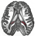
A view showing the ventricles inside the brain.
-
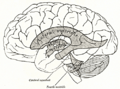
A diagram showing where the ventricles are located compared to the brain's surface.
-
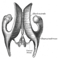
A drawing of the ventricular cavities, seen from above.
-
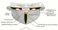
A view of the ventricles and the choroid plexus.
-
The lateral ventricles and other brain structures, shown in a transparent brain view.


See also
 In Spanish: Ventrículo cerebral para niños
- Blood–brain barrier
- Circumventricular organs
