
X-ray facts for kids

X-rays are a special type of electromagnetic radiation. Think of them as invisible waves, similar to light waves, but with much more energy. They have a very short wavelength, which means they can do things visible light can't. X-rays are part of a bigger family called the electromagnetic spectrum, which includes everything from radio waves to gamma rays.
Most X-rays have wavelengths between 0.01 and 10 nanometres. To give you an idea, a nanometre is one billionth of a meter! This short wavelength gives them a lot of energy, which is why they are so powerful.
Contents
How X-rays See Inside You
X-rays are amazing because they can pass through many solid things. This is why doctors use them to take pictures inside your body, especially to see your bones. Sometimes, people even call these pictures "X-rays" instead of the radiation itself.
Why Bones Show Up
When an X-ray picture is taken, it works because different parts of your body block the X-rays differently. Your bones are very dense, meaning they are packed tightly with material. Because of this, X-rays can't easily pass through them. Instead, the X-rays are either soaked up (absorbed) or bounced away (scattered) by the bones. This makes the bones show up as white or light areas on the X-ray picture.
What X-rays Don't Show
On the other hand, your skin and muscles are not as dense. X-rays can pass through these tissues much more easily without being absorbed too much. That's why you can't see your skin or muscles clearly on a standard X-ray. To see things like tumors or soft tissues, doctors use other tools like magnetic resonance imaging (MRI).
3D X-ray Pictures
A special machine called a computed tomography scanner (CT scanner) combines an X-ray machine with a computer. This allows it to create a three-dimensional (3D) picture of the inside of your body. CT scans can show more than just bones, giving doctors a more detailed view.
How X-rays Are Made
X-rays are created when very fast-moving electrons hit a piece of metal. These electrons make tiny packets of energy called photons, which are the X-rays themselves. These photons can move atoms and cause chemical changes in your body.
Different Types of X-rays
The effects of X-rays depend on how much energy they have:
- "Soft" X-rays: These have lower energy. They are often used for making pictures of people.
- "Hard" X-rays: These have higher energy. They are powerful enough to be used in radiation therapy to treat cancer.
X-rays and Your Health
There are tiny amounts of X-rays all around us in the air. Like other types of energy in the air, X-rays can affect living cells.
Safety with X-rays
It's important to know that being exposed to a lot of X-rays for a long time can be risky. High doses can even increase the chance of getting cancer. However, medical X-rays are carefully controlled to use the smallest amount of radiation needed.
X-rays as Medicine
Even though high doses can be harmful, X-rays are also used to help people. Cancer cells are often more sensitive to radiation than healthy cells. This means that doctors can sometimes use X-rays to kill cancer cells while trying to protect the healthy ones.
Related pages
Images for kids
-
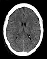
Head CT scan (transverse plane) slice – a modern application of medical radiography
-
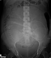
Abdominal radiograph of a pregnant woman, a procedure that should be performed only after proper assessment of benefit versus risk
-
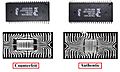
Using X-ray for inspection and quality control: the differences in the structures of the die and bond wires reveal the left chip to be counterfeit.
-
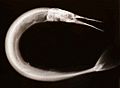
X-ray fine art photography of needlefish by Peter Dazeley
-

Wilhelm Röntgen
-
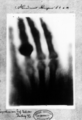
Hand mit Ringen (Hand with Rings): print of Wilhelm Röntgen's first "medical" X-ray, of his wife's hand, taken on 22 December 1895 and presented to Ludwig Zehnder of the Physik Institut, University of Freiburg, on 1 January 1896
-
Pulyui's X-ray images
-

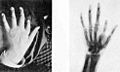
Taking an X-ray image with early Crookes tube apparatus, late 1800s. The Crookes tube is visible in center. The standing man is viewing his hand with a fluoroscope screen. The seated man is taking a radiograph of his hand by placing it on a photographic plate. No precautions against radiation exposure are taken; its hazards were not known at the time.
-
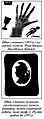
1896 plaque published in "Nouvelle Iconographie de la Salpetrière", a medical journal. In the left a hand deformity, in the right same hand seen using radiography. The authors designated the technique as Röntgen photography.
-
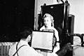
A patient being examined with a thoracic fluoroscope in 1940, which displayed continuous moving images. This image was used to argue that radiation exposure during the X-ray procedure would be negligible.
-

Chandra's image of the galaxy cluster Abell 2125 reveals a complex of several massive multimillion-degree-Celsius gas clouds in the process of merging.
-
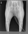
Dog hip xray posterior view
-
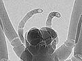
Phase-contrast x-ray image of spider


See also
 In Spanish: Rayos X para niños
