
Alzheimer's disease facts for kids

Alzheimer's Disease (AD) is a brain disease that slowly damages brain cells. Right now, there is no cure for Alzheimer's disease. Over time, the problems caused by the disease become more serious. Many people die because of Alzheimer's. This disease mainly affects parts of the brain that control memory, language, and thinking skills. Alzheimer's is the most common type of dementia in older people.
Symptoms of Alzheimer's usually appear after age 65. However, changes in the brain that cause Alzheimer's can start many years before any symptoms show up. Even though it affects older people, Alzheimer's is not a normal part of aging.
There is no cure for Alzheimer's yet. But there are treatments that can help with the symptoms. These treatments can make the symptoms less severe. Some treatments can also slow down the disease. This means the damage to the brain happens more slowly. Also, certain healthy habits might help delay the start of the disease.
Scientists don't know exactly what causes Alzheimer's disease. But there are several risk factors that can make a person more likely to get it. Some of these risks are genetic. Changes in four different genes have been found that increase the risk.
Alzheimer's disease was named after Alois Alzheimer. He was a German doctor who first described the disease. This was after he studied a patient named Auguste Deter in 1906. The disease was officially named Alzheimer's disease in 1910.
Contents
What Happens in the Brain?


In the brains of people with Alzheimer's disease, doctors find two main problems. These are neurofibrillary tangles (or 'tangles') and senile plaques (or 'plaques').
Tangles are made of a protein called tau. Plaques are mostly made from another protein called beta-amyloid.
- Tangles: Inside healthy brain cells (neurons), there are tiny structures called microtubules. These are like tiny roads that help the cell work and communicate. A protein called tau usually holds these microtubules together. In Alzheimer's, the tau protein changes and breaks away. It then forms twisted clumps, or tangles, inside the neurons. When this happens, the microtubules fall apart. This stops the brain cells from working properly and eventually kills them.
- Plaques: Plaques form outside the brain cells. They start with a protein called amyloid precursor protein (APP). This protein is part of the outer covering of brain cells. In Alzheimer's, special chemicals in the brain cut APP into small pieces. Some of these pieces are called beta-amyloid. These beta-amyloid pieces then stick together. They form sticky clumps, or plaques, in the spaces between brain cells. These plaques can block signals between brain cells and harm them.
Scientists are still studying if tangles and plaques cause Alzheimer's. Or if they are a result of the disease.
How Alzheimer's Spreads in the Brain


Alzheimer's disease usually starts in a small area deep inside the brain. This area is called the "transentorhinal region." Brain cells start to die here first.
Then, the disease spreads to a nearby area called the "entorhinal cortex" (EC). The EC is like a main station for brain signals. It connects to many parts of the brain that handle memory and movement.

The EC is very important for communication between the hippocampus and the neocortex. The neocortex is the outer part of the brain. It helps us with higher functions. These include how we understand what we see, hear, smell, taste, and touch. It also helps us move, think, and use language.
Next, the disease spreads to the hippocampus. The hippocampus is part of the brain's limbic system. It helps us form new memories, organize them, and store them. It also connects emotions and senses (like smells or sounds) to specific memories. For example, a certain smell might bring back a memory.
The hippocampus sends memories to other parts of the brain for long-term storage. It also helps us remember things later. For example, an adult trying to remember a classmate's name from kindergarten.
Besides memory, the hippocampus also helps with emotions, finding our way around, and knowing where we are in space. For example, knowing your way around your bedroom even with the lights off. There are two parts of the hippocampus, one on the left side of the brain and one on the right.
How Doctors Find Alzheimer's

Red Blue Green Purple Orange
Purple Orange Green Blue Red
Blue Orange Purple Green Red
Purple Green Red Blue Orange
This test measures how well a person pays attention.
It's easier to name the colors of the first set of words because the color matches the word. In the second set, the colors don't match the words. So, you have to pay more attention.
People with early Alzheimer's often have trouble with this test.Before Symptoms Appear New research tools can now detect early signs of Alzheimer's disease. These signs appear even before a person has any symptoms. For example, special brain scans can show if amyloid beta (Aβ) is building up in the brain. This build-up is linked to changes in brain structure and function. These changes are similar to what is seen in people with memory problems or full Alzheimer's.
These very early changes in the brain can happen many years, even decades, before someone is diagnosed. Not everyone with these early changes will get the disease. But it does put them at higher risk. Even though there's no cure, new treatments are being developed. These treatments might work best in the very first stages of the disease.
Scientists around the world are working together to study these early stages. This research helps them understand why some people develop Alzheimer's and others don't.
Early Stages It can be hard to diagnose Alzheimer's in its very early stages. More than 100 other conditions can look like the disease. For people with only mild memory problems, it's difficult to be sure it's Alzheimer's. Doctors need better ways to look inside the brain to make a clear diagnosis.
History of Alzheimer's Disease


He studied Auguste Deter when she was alive. He also looked at her brain under a microscope after she died.

In 1901, a 51-year-old woman named Auguste Deter was admitted to a hospital in Frankfurt am Main, Germany. This hospital was nicknamed "Castle of the Insane." She had been married and lived a normal life. But eight months before, she started having problems with memory, language, and thinking. She also became confused and had hallucinations.
A doctor named Alois Alzheimer (1864–1915) studied her case. He was very interested because of her age. At that time, memory loss in older people was known. But it usually started in their mid-sixties. Auguste's case was unusual because her problems started quickly and at a younger age.
During one of his exams, Dr. Alzheimer asked Ms. Deter to write her name. She couldn't do it. She said, "I have lost myself, so to speak."
Ms. Deter died on April 8, 1906. Her brain was sent to Dr. Alzheimer for study. He looked at samples of her brain under a microscope. He saw the neurofibrillary tangles and beta-amyloid plaques. These are the two main features of the disease.
On November 3, 1906, Dr. Alzheimer shared his findings about Auguste's case. He published his research in 1907. In 1910, his colleague, Emil Kraepelin, named the disease 'Alzheimer's disease'. Auguste Deter was 55 when she died. Her case is now known as an example of Early-onset Alzheimer's disease. This is when the disease starts at a younger age.
Famous People with Alzheimer's

Anyone can get Alzheimer's disease, no matter if they are rich or poor, famous or not. Some well-known people who have had Alzheimer's disease include:
- Former U.S. President Ronald Reagan
- Irish writer Iris Murdoch
- Footballer Ferenc Puskás
- Former Prime Ministers Harold Wilson (United Kingdom) and Adolfo Suárez (Spain)
- Novelist Terry Pratchett
- Politician and activist Sargent Shriver
- Nobel Prize winner in Physics, Charles K. Kao
- Nobel Prize writer Gabriel García Márquez
These famous cases show that Alzheimer's can affect anyone.
Images for kids
-
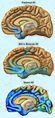
Stages of atrophy (shrinking) in Alzheimer's.
-
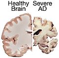
A normal brain on the left and a late-stage Alzheimer's brain on the right.
-
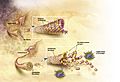
In Alzheimer's disease, changes in tau protein lead to the breakdown of tiny structures in brain cells.
-
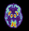
PET scan of the brain of a person with Alzheimer's disease showing less activity in the temporal lobe.
-
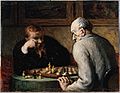
Activities like playing chess or being social might lower the risk of Alzheimer's.
-

A model of donepezil, a medicine used to help with Alzheimer's symptoms.
-
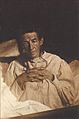
Alois Alzheimer's patient Auguste Deter in 1902. Her case was the first to be described.
.jpg)


See also
 In Spanish: Enfermedad de Alzheimer para niños
