
Electron crystallography facts for kids
Electron crystallography is a cool way to figure out how tiny atoms are arranged inside solid materials. It uses a special powerful microscope called a transmission electron microscope (TEM).
This method is super helpful in many situations where another method, called X-ray crystallography, can't be used. X-ray crystallography usually needs large, perfect crystals to work properly.
Electron crystallography is great for studying things like proteins, which often form flat, 2-dimensional layers (like sheets or spirals called helices), or tiny shapes like the outer shells of viruses (called capsids). It can also look at proteins that are just floating around. Electrons work well in these cases because they interact much more strongly with atoms than X-rays do.

Contents
What is Electron Crystallography?
Electron crystallography is a scientific method. It helps scientists see how atoms are arranged in solid materials. It uses a special tool called a transmission electron microscope (TEM).
This method was first developed by a scientist named Aaron Klug. He even won the Nobel Prize in Chemistry in 1982 for his work. He also studied virus structures and how cells make proteins.
Discoveries with Electron Crystallography
The first protein structure solved using electron crystallography was bacteriorhodopsin. This happened in 1990.
Since then, scientists have used this method to study many other important structures. These include the light-harvesting complex, which helps plants capture sunlight. They also studied the bacterial flagellum, which is like a tiny motor that helps bacteria move.
Related pages
Images for kids
-
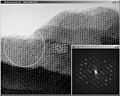
Electron microscopy image of an inorganic tantalum oxide. The inset shows its Fourier transform. Notice how the appearance changes from the thin upper region to the thicker lower region. The unit cell of this compound is about 15 by 25 Ångström. It is outlined at the center of the figure, inside the result from image processing, where the symmetry has been taken into account. The black dots show clearly all the tantalum atoms. The diffraction extends to 6 orders along the 15 Å direction and 10 orders in the perpendicular direction. Thus the resolution of the EM image is 2.5 Å (15/6 or 25/10). This calculated Fourier transform contains both amplitudes (as seen) and phases (not displayed).

