
Mitosis facts for kids

Mitosis is a super important process where a cell divides into two identical cells. Think of it like a cell making an exact copy of itself! This process is a key part of the cell cycle, which is how cells grow and reproduce.
Before a cell can divide, it needs to make a perfect copy of its genetic information. This information is stored in structures called chromosomes. Each chromosome contains DNA, which is like the cell's instruction manual. When a cell gets ready for mitosis, these chromosomes make exact copies of themselves. Each copied chromosome then looks like two identical halves, called sister chromatids, joined together in the middle by a centromere.
Mitosis happens in almost all the cells in your body that need to divide, like skin cells or muscle cells. However, it doesn't happen in the cells that create sperm or eggs (these are called gametes or sex cells). Those special cells use a different kind of division called meiosis.
Contents
How Cells Divide: The Stages of Mitosis

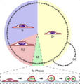
Mitosis happens in several clear stages. Each stage has specific things that happen inside the cell to make sure the chromosomes are divided perfectly. These stages are called prophase, prometaphase, metaphase, anaphase, and telophase.
Prophase: Getting Ready to Divide

During prophase, the chromosomes inside the cell's nucleus start to coil up and become thicker. This makes them easier to see under a light microscope. Also, special structures called centrioles move to opposite sides of the nucleus. These centrioles help form spindle fibers, which are like tiny ropes that will pull the chromosomes apart.
Prometaphase: The Nucleus Opens Up

In prometaphase, the membrane around the nucleus, called the nuclear envelope, breaks down. This means the sister chromatids are now free inside the cell. A special protein structure called a kinetochore forms on each centromere (where the sister chromatids are joined). The spindle fibers, which are long, thin proteins, reach out from opposite ends of the cell and attach to these kinetochores.
Metaphase: Lining Up in the Middle

During metaphase, the spindle fibers pull and push the sister chromatids until they all line up perfectly in the very center of the cell. This imaginary line in the middle is often called the "metaphase plate" or the cell's equator. It's like a tug-of-war where everyone ends up in the middle, ready for the big split.
Anaphase: Pulling Apart

Anaphase is when the sister chromatids finally separate! The spindle fibers attached to the kinetochores shorten, pulling the individual chromatids (now considered full chromosomes again) away from the cell's center and towards opposite ends, or "poles," of the cell. Other spindle fibers that are not attached to chromosomes actually get longer, helping to push the ends of the cell further apart. The cell starts to stretch out, getting ready to divide into two.
Telophase: Two New Nuclei Form
Telophase is the last stage of mitosis. Now, there's a complete set of identical chromosomes at each end of the stretched-out cell. The spindle fibers start to disappear. A new nuclear membrane forms around each set of chromosomes, creating two brand new nuclei. Inside each new nucleus, a nucleolus (a small, dense spot) reappears, and the chromosomes uncoil, becoming less visible.
Cytokinesis: The Cell Splits in Two
Even though it's super important for cell division, cytokinesis isn't technically part of mitosis itself. It's the final step where the entire cell physically splits into two separate "daughter" cells. This usually happens at the same time as anaphase and telophase.
In animal cells, the cell membrane pinches inward, forming a "cleavage furrow" that gets deeper and deeper until the cell completely divides. In plant cells, it's a bit different. Instead of pinching, a new cell wall forms in the middle, creating a "cell plate" that grows until it divides the cell into two.
After cytokinesis, the two new daughter cells are ready to start their own lives. They go back into the "interphase" stage of the cell cycle, where they grow and prepare for their own division, repeating the cycle all over again!
Images for kids
-
Label-free live cell imaging of Mesenchymal Stem Cells undergoing mitosis
-
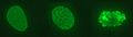
Condensing chromosomes. Interphase nucleus (left), condensing chromosomes (middle) and condensed chromosomes (right).
-
A cell in late metaphase. All chromosomes (blue) but one have arrived at the metaphase plate.
-
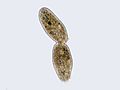
Cilliate undergoing cytokinesis, with the cleavage furrow being clearly visible
-
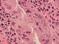
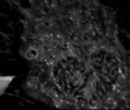
An abnormal (tripolar) mitosis (12 o'clock position) in a precancerous lesion of the stomach (H&E stain)
-

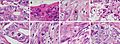
Mitosis appearances in breast cancer
-
Cell shape changes through mitosis for a typical animal cell cultured on a flat surface. The cell undergoes mitotic cell rounding during spindle assembly and then divides via cytokinesis. The actomyosin cortex is depicted in red, DNA/chromosomes purple, microtubules green, and membrane and retraction fibers in black. Rounding also occurs in live tissue, as described in the text.
-
Normal and atypical forms of mitosis in cancer cells. A, normal mitosis; B, chromatin bridge; C, multipolar mitosis; D, ring mitosis; E, dispersed mitosis; F, asymmetrical mitosis; G, lag-type mitosis; and H, micronuclei. H&E stain.
-
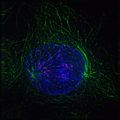
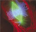
Early prophase: Polar microtubules, shown as green strands, have established a matrix around the currently intact nucleus, with the condensing chromosomes in blue. The red nodules are the centromeres.


See also
 In Spanish: Mitosis para niños
