
DNA facts for kids


DNA, which is short for deoxyribonucleic acid, is a special molecule. It holds the instructions, or "genetic code," for all living things. This includes animals, plants, tiny protists, archaea, and bacteria.
Every cell in an organism has DNA. This DNA tells the cells exactly what proteins to create. Most of these proteins are enzymes, which help with many jobs in the cell. Children get their DNA from their parents. This is why kids often look like their parents, sharing traits like hair or eye color. A person's DNA is a mix of DNA from both their mother and father.
Some parts of an organism's DNA are called "non-coding DNA". These parts do not contain instructions for making proteins. However, some non-coding DNA is used to make special non-coding RNA molecules. These include transfer RNA and ribosomal RNA, which are important for making proteins. Other non-coding parts might not be used at all, or their purpose is still a mystery. The amount of non-coding DNA can be very different between species. For example, over 98% of human DNA is non-coding, but only about 2% of a typical bacterial genome is non-coding.
Viruses use either DNA or RNA to infect other organisms. Most DNA viruses copy their DNA inside the cell's nucleus. RNA viruses, however, usually copy their RNA in the cytoplasm, which is the jelly-like substance filling the cell.
Contents
What is DNA's Structure?
DNA has a unique shape called a double helix. Imagine a ladder that has been twisted into a spiral. Each step of this twisted ladder is made of two matching parts called nucleotides.
DNA's Building Blocks: Nucleotides
A nucleotide is a small molecule with three main parts:
- A type of sugar called deoxyribose, which has five carbon atoms.
- A phosphate group, made from phosphorus and oxygen.
- A nitrogenous base.
There are four different types of nucleotides that make up DNA:
The "rungs" of the DNA ladder are formed by two bases joining together. One base comes from each side of the ladder. These bases connect in a very specific way: 'A' always pairs with 'T', and 'C' always pairs with 'G'. These pairs are held together by weak connections called hydrogen bonds.
Adenine (A) and thymine (T) pair up because they can form two hydrogen bonds. Cytosine (C) and guanine (G) pair up by forming three hydrogen bonds. Even though the bases always pair in these specific ways, the order of the pairs can change. For example, you can have A-T or T-A, and C-G or G-C. This changing order allows DNA to create "codes" using these "letters" (the bases). These codes carry the important messages that tell a cell what to do.
DNA and Proteins: Chromatin
Inside the cell, DNA is tightly wrapped around special proteins called histones. This combination of DNA and proteins is called chromatin. This wrapping helps control which genes are active or inactive. Genes are like switches that turn on and off during development and cell activities. This control is a big part of how cells work.
How DNA Makes Copies
When DNA makes a copy of itself, it's called DNA replication. To do this, the hydrogen bonds holding the base pairs together break. The DNA molecule then splits down the middle, like unzipping a zipper. This creates two single strands. New strands are then built by matching the correct bases (A with T, and G with C) to each of the separated strands.
First, an enzyme called DNA helicase acts like a tiny unzipper. It breaks the hydrogen bonds, splitting the DNA molecule into two separate pieces. After that, another molecule called DNA polymerase comes along. It builds a new matching strand for each of the original separated strands. This means that each new DNA molecule is made of one old strand and one brand new strand.
DNA Copying Mistakes: Mutations
Sometimes, when DNA is copied, small mistakes happen. These mistakes are called mutations. There are a few main types of mutations:
- Deletion: One or more bases are accidentally left out.
- Substitution: One or more bases are swapped for a different base.
- Insertion: One or more extra bases are added into the sequence.
- Duplication: A sequence of base pairs is accidentally repeated.
Mutations can also be grouped by how they affect proteins or an organism's health. Some mutations can be harmful, some have no effect, and some can even be helpful. Sometimes, a mutation can be very bad, causing the new protein to not work at all, which can even cause an embryo to die. But on the other hand, mutations are a key part of evolution. If a new version of a protein works better for an organism, it can help that organism survive and thrive.
How Proteins Are Made
A section of DNA that holds the instructions for making a specific protein is called a gene. Each gene contains the code for at least one polypeptide, which is a chain of amino acids. Proteins build structures in the cell and also form enzymes, which do most of the cell's work. To make a protein for a certain job, the right amino acids must be linked together in the correct order.
Proteins are built by tiny machines inside the cell called ribosomes. Ribosomes are found in the main part of the cell, but DNA stays safely inside the cell's nucleus. Since DNA cannot leave the nucleus, the cell makes a copy of the DNA sequence using RNA. This RNA copy is smaller and can fit through tiny holes, called pores, in the nucleus's outer layer to get out into the cell.
The instructions encoded in DNA are first copied into messenger RNA (mRNA) by proteins like RNA polymerase. This mRNA then acts as a template for the ribosome to build the protein. Ribosomes read "words" called codons, which are made of three base pairs. Each codon tells the ribosome which amino acid to add next. The ribosome moves along the mRNA, reading the code and building the protein. Another type of RNA, called tRNA, helps to bring the correct amino acid to each codon.
History of DNA Discovery

The first time DNA was taken out of cells was in 1869. A Swiss physician named Friedrich Miescher did this while studying bacteria from surgical bandages. He found this molecule in the cell's nucleus and named it nuclein.
In 1928, Frederick Griffith found something amazing. He discovered that traits from one type of bacteria could be transferred to another type. This was the first strong hint that DNA carried genetic information.
Later, in 1943, the Avery–MacLeod–McCarty experiment showed that DNA was indeed the "transforming principle." This meant DNA was responsible for passing on traits.
DNA's role in heredity was fully confirmed in 1952. Alfred Hershey and Martha Chase showed that DNA was the genetic material of a virus that infects bacteria.
In the 1950s, Erwin Chargaff made an important discovery. He found that in a DNA molecule, the amount of adenine (A) was always about equal to the amount of thymine (T). He also found that the amount of guanine (G) was about equal to the amount of cytosine (C). These findings are known as Chargaff's rules.
In 1953, James D. Watson and Francis Crick proposed the now-famous double-helix model of DNA structure. They published their idea in the journal Nature. Their model was based on an X-ray diffraction image, known as "Photo 51." This image was taken by Rosalind Franklin and Raymond Gosling in 1952.
Other scientists also published evidence supporting the Watson and Crick model. In 1962, Watson, Crick, and Maurice Wilkins received the Nobel Prize in Physiology or Medicine. Rosalind Franklin had passed away in 1958, and Nobel Prizes are only given to living people. There is still much discussion about who should get credit for the discovery.
In 1957, Crick explained how DNA, RNA, and proteins work together. This idea is called the central dogma of molecular biology.
How DNA copies itself was fully understood in 1958, thanks to the Meselson–Stahl experiment. More work by Crick and others showed that the genetic code uses "words" made of three bases, called codons. These discoveries marked the beginning of molecular biology.
DNA and Privacy
In some places, like the United States, police have used DNA from public family tree databases to help solve old, unsolved cases. This practice has led to discussions about privacy concerns.
Related pages
Images for kids
-
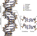
The structure of the DNA double helix. The atoms in the structure are colour-coded by element and the detailed structures of two base pairs are shown in the bottom right.
-
animated version]]).
-
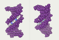
DNA major and minor grooves. The latter is a binding site for the Hoechst stain dye 33258.
-
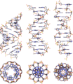
From left to right, the structures of A, B and Z DNA
-
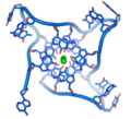
DNA quadruplex formed by telomere repeats. The looped conformation of the DNA backbone is very different from the typical DNA helix. The green spheres in the center represent potassium ions.
-
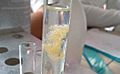
Impure DNA extracted from an orange
-
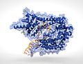
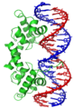
T7 RNA polymerase (blue) producing an mRNA (green) from a DNA template (orange)
-
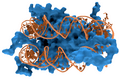
Interaction of DNA (in orange) with histones (in blue). These proteins' basic amino acids bind to the acidic phosphate groups on DNA.
-
The lambda repressor helix-turn-helix transcription factor bound to its DNA target
-
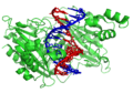
The restriction enzyme EcoRV (green) in a complex with its substrate DNA
-
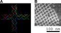
The DNA structure at left (schematic shown) will self-assemble into the structure visualized by atomic microscopy at right. DNA nanotechnology is the field that seeks to design nanoscale structures using the molecular recognition properties of DNA molecules.
-
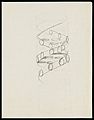
Pencil sketch of the DNA double helix by Francis Crick in 1953


.jpg)


See also
 In Spanish: Ácido desoxirribonucleico para niños
