
Kinetoplast facts for kids
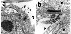
A kinetoplast is a special network of circular DNA found inside the mitochondrion of certain tiny living things. Think of it like a tiny, organized bundle of genetic instructions. This DNA, called kDNA, holds many copies of the cell's mitochondrial genome. Kinetoplasts usually look like a flat disk, but they can have other shapes too. You can only find kinetoplasts in a group of single-celled organisms called Kinetoplastida. These organisms are part of a larger group called Excavata. The different shapes of kinetoplasts can help scientists understand how these organisms are related to each other. A kinetoplast is often located right next to the organism's flagella (a tail-like structure used for movement) and its basal body (the anchor for the flagellum). This closeness suggests it's connected to the cell's internal support system, called the cytoskeleton.
Contents
Kinetoplasts in Tiny Organisms
Many interesting organisms have kinetoplasts. For example, trypanosomes are tiny, flagellated (meaning they have flagella) single-celled creatures. In these organisms, the kinetoplast is a dense clump of DNA inside their mitochondrion.
One famous example is Trypanosoma brucei. This parasite causes a serious illness called African trypanosomiasis, also known as African sleeping sickness. Scientists can easily see its kinetoplast when they stain samples with special dyes like DAPI, which makes DNA glow under a microscope.
Another parasite, Trypanosoma cruzi, causes Chagas disease. This disease mostly affects people in Central and South America and is spread by insects often called "kissing bugs." Even though African sleeping sickness is very serious, the kinetoplast in T. cruzi is much larger than the one in T. brucei.
There's also Trypanosoma equiperdum, which causes a disease called dourine in horses. This is a unique type of trypanosome infection that spreads between horses. The kinetoplasts in T. equiperdum are special because all their small DNA circles (called minicircles) have the exact same genetic code.
How Kinetoplasts are Built
The kinetoplast contains two main types of circular DNA: maxicircles and minicircles.
- Maxicircles are larger, about 20 to 40 thousand base pairs long. There are usually a few dozen of these in each kinetoplast. They hold the instructions for making the proteins needed by the mitochondrion.
- Minicircles are much smaller, only about 500 to 1000 base pairs long. There are thousands of these in each kinetoplast. Their main job is to produce special guide RNA (gRNA). This gRNA helps to "decode" the information stored in the maxicircles, often by adding or removing tiny parts of the genetic code.
Imagine these maxicircles and minicircles are like tiny rings. They are all linked together, forming a flat network that looks a bit like chain mail. When the organism needs to make a copy of this network, these rings have to be temporarily unlinked from the parent kinetoplast and then reconnected in the new, daughter kinetoplast. This unique way of copying DNA could give scientists ideas for new medicines.
The kinetoplast structure that scientists know best comes from Crithidia fasciculata. In this organism, the kinetoplast is a disk made of linked maxicircles and minicircles. Most of these DNA circles are not tightly coiled. Around the edge of this DNA disk, there are two protein groups, located opposite each other. These groups are important for copying the minicircles. Scientists have studied the network of DNA in C. fasciculata to understand how all these circles are connected. Early studies suggested a honeycomb lattice structure, but more recent detailed imaging shows it's actually quite disordered, though each minicircle is still linked to about three others.
Different Kinds of Kinetoplasts
Scientists have found different types of kinetoplast networks, which vary in how their kDNA is arranged and where it's located. These differences help us understand the evolutionary journey of kinetoplastids.
- A pro-kDNA kinetoplast looks like a bundle. It's found inside the mitochondrion, close to the flagellum's anchor point. Unlike the usual kinetoplast, pro-kDNA has very few linked circles, and its maxicircles and minicircles are relaxed, not tightly coiled. You can find pro-kDNA in organisms like Bodo saltans and Rhynchomonas nasuta.
- A poly-kDNA kinetoplast is similar to pro-kDNA in that it has few linked circles and no tight coiling. The special thing about poly-kDNA is that it's not one single bundle. Instead, it's spread out in several separate spots (called foci) throughout the mitochondrion. Dimastigella trypaniformis, a tiny organism living in the gut of termites, has poly-kDNA.
- A pan-kDNA kinetoplast also has fewer linked circles, but its minicircles are tightly coiled. Pan-kDNA kinetoplasts fill most of the mitochondrion and are not limited to separate spots. This type is seen in Cryptobia helicis, a parasite found in snails, and Bodo caudatus.
- A mega-kDNA kinetoplast is spread evenly throughout the mitochondrion. It's unique because it doesn't have minicircles. Instead, the DNA sequences that are similar to minicircles are joined together into much longer molecules, about 200 thousand base pairs long. Mega-kDNA has been observed in parasites like Trypanoplasme borreli (a fish parasite) and Jarrellia sp. (a whale parasite).
These different kDNA structures show how kinetoplastids have changed over time. Pan-kDNA, which looks a lot like a simple DNA plasmid, might be the original form of kDNA from which others evolved.
How Kinetoplasts Make Copies

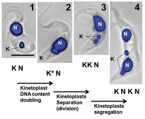
The kinetoplast copies itself at the same time the nearby flagellum duplicates, and just prior to the cell's main DNA (nuclear DNA) is copied. In a typical Crithidia fasciculata kinetoplast, the copying process starts when special proteins called topoisomerase II unhook the minicircles from the network.
These free minicircles move into a special area between the kinetoplast and the mitochondrion's outer wall. After they are copied, the minicircles travel to two protein groups located on opposite sides of the kinetoplast. These groups contain several proteins that help with copying, including those that fix any breaks in the newly made minicircles.
This copying happens one minicircle at a time. Only a few minicircles are unhooked at any moment. To keep track of which minicircles have been copied, a small gap is left in the new minicircles when they rejoin the network. This gap marks them as already copied. Minicircles that haven't been copied yet are still fully closed circles. After copying, each new minicircle attaches to the kinetoplast network near the protein groups, and the gaps are partly fixed.

As more minicircles are copied, the entire kDNA network rotates around its center. This rotation helps prevent too many new minicircles from building up in one place. Scientists believe this spinning is connected to the copying of the nearby flagellum. The new flagellum's anchor point also rotates around the old one in a similar way. By rotating, the new minicircles are added in a spiral pattern and move towards the center of the disk as more minicircles are unhooked and copied.
While we know a lot about minicircle copying, the exact details for maxicircle kDNA are still being studied. However, scientists have observed a structure called a nabelschnur (which means "umbilical cord" in German). This structure tethers, or connects, the two new kDNA networks together before they finally break apart during cell division. Studies show that the nabelschnur contains maxicircle kDNA.
Kinetoplast copying happens in five main stages, which are linked to how the nearby flagellum also copies itself:
- Stage I: The kinetoplast is not yet copying itself. It has no special protein groups and is next to a single flagellum anchor.
- Stage II: The kinetoplast starts to show the protein groups. The flagellum anchor begins to copy, and so does the kinetoplast. This makes the kinetoplast look somewhat dome-shaped.
- Stage III: The new flagellum starts to separate, and the kinetoplast begins to look like it has two lobes (two rounded parts).
- Stage IV: The kinetoplasts appear as two separate disks, but they are still connected by the nabelschnur.
- Stage V: The two daughter kinetoplasts completely separate when the nabelschnur breaks. Their structure then looks just like it did in Stage I.
Kinetoplasts and DNA Repair
Trypanosoma cruzi has an amazing ability to fix its DNA. When this parasite infects a host, the host's body produces harmful substances called reactive oxygen species. These can damage the parasite's DNA, both in its main genome and in its kinetoplast.
However, T. cruzi has a special protein called DNA polymerase beta. This protein helps to remove and repair these oxidative DNA damages through a process called base excision repair. It seems that DNA polymerase beta is especially active during kinetoplast DNA replication. This helps to fix any DNA damage caused by stress in this important organelle while it's making copies.
