
Cranial nerve facts for kids

A cranial nerve is a special nerve that connects straight to your brain or brainstem. This is different from spinal nerves, which connect to your spinal cord. Cranial nerves help your brain talk directly to parts of your body, mostly your head and neck.
Humans have twelve pairs of cranial nerves. They are numbered using Roman numerals I to XII. This numbering is based on their order from the front of your brain to the back. Each nerve pair is found on both sides of your central nervous system.
Contents
- What Cranial Nerves Do
- How Nerves Leave the Brain
- Related pages
- Images for kids
- See also
What Cranial Nerves Do
Cranial nerves help you feel things and move parts of your head and neck. They let you experience sensations like taste, vision, smell, balance, hearing, touch, and temperature. They also control many muscles.
Smell (I)
The olfactory nerve (CN I) helps you smell. It sends information about smells from your nose to your brain. This nerve is a sensory nerve, meaning it takes information from the outside world and sends it to your central nervous system.
How the Smell Nerve Works
The olfactory nerve is the shortest cranial nerve. It is made of many tiny neurons that work together. These neurons start in your nasal cavity (inside your nose). They reach up through the roof of your nasal cavity to connect to a part of your brain called the olfactory bulb.
When you breathe in, tiny smell molecules enter your nasal cavity. The olfactory neurons detect these molecules. They turn the smell signals into electrical signals. These electrical signals travel up the neurons to the olfactory bulb. From there, the signals go to different parts of your brain. For example, some signals go to the frontal lobe, which helps you figure out what you are smelling.
If this nerve gets damaged, you might lose your sense of smell. This is called anosmia.

Vision (II)
The optic nerve (CN II) sends visual information from your retina (at the back of your eye) to your brain. This allows you to see.
Eye Movement (III, IV, VI)
- The oculomotor nerve (CN III) controls most of the muscles that move your eye. It also helps control your eyelids and the size of your pupil.
- The trochlear nerve (CN IV) controls one specific eye muscle.
- The abducens nerve (CN VI) controls another specific eye muscle.
Facial Sensation and Jaw Movement (V)
The trigeminal nerve (CN V) is named "trigeminal" because it has three main parts. Together, these parts help you feel sensations in your face. They also control big facial movements like biting and chewing.
Facial Expression (VII)
The facial nerve (CN VII) controls the muscles that let you make different facial expressions. It also helps carry taste sensations from the back of your tongue and mouth.
Hearing and Balance (VIII)
The vestibulocochlear nerve (CN VIII) sends information about sound and balance from your inner ear to your brain. This helps you hear and keep your balance.
Oral Sensation, Taste, and Salivation (IX)
The glossopharyngeal nerve (CN IX) carries many different types of sensory and motor information. It helps with sensations in your mouth, taste, and making saliva.
Control of Heart and Digestion (X)
The vagus nerve (CN X) helps control your heart and digestive system. It is the longest nerve in your autonomic nervous system.
Shoulder Elevation and Head-Turning (XI)
The accessory nerve (CN XI) controls two important muscles: the sternocleidomastoid and trapezius muscles. These muscles help you lift your shoulders and turn your head.
Tongue Movement (XII)
The hypoglossal nerve (CN XII) helps control your tongue movements. These movements are important for speaking and swallowing food.
How Nerves Leave the Brain
After leaving the brain, cranial nerves travel inside your skull. To reach their destinations, some nerves pass through small holes in the skull called foramina. Other nerves travel through longer pathways made of bone, called bony canals. Sometimes, more than one cranial nerve, and even blood vessels, can pass through the same hole or canal.
Related pages
Images for kids
-
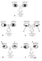
The oculomotor (III), troclear (IV) and abducens (VI) nerves supply the muscle of the eye. Damage will affect the movement of the eye in various ways, shown here.
-

The facial nerve (VII) supplies the muscles of facial expression. Damage to the nerve causes a lack of muscle tone on the affected side, as can be seen on the right side of the face here.
-
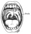
A damaged glossopharyngeal nerve (IX) may cause the uvula to deviate to the affected side.
-
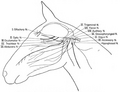
The cranial nerves in the horse.
-
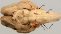
Ventral view of a sheep's brain. The exits of the various cranial nerves are marked with red.


See also
 In Spanish: Pares craneales para niños
