
Hemoglobin facts for kids

Hemoglobin is a super important protein found in your red blood cells. Think of it as a tiny delivery truck that carries oxygen from your lungs to every part of your body! It helps your body get the oxygen it needs to work properly.
Hemoglobin has a special part called heme. This heme contains iron, which is what actually grabs onto the oxygen. Doctors can check your hemoglobin levels with a simple blood test. These levels are usually measured in grams per deciliter (g/dL).
If your hemoglobin levels are too low, it can make you feel tired, weak, or dizzy. This might be a sign of a condition like anemia. A doctor can find out why your levels are low and help you feel better.
What is Hemoglobin Made Of?
The most common type of hemoglobin in people has four small parts, called subunits. Each subunit is a protein called globin, and inside each globin is a heme group.
So, a complete hemoglobin molecule has four globin chains, four heme molecules, and four iron atoms. When hemoglobin is in your lungs, it picks up oxygen. Then, it carries this oxygen to all the other parts of your body that need it.
Scientists worked for many years to understand how hemoglobin is built. Max Perutz and John Kendrew were two important scientists who helped figure out its structure. They first studied a similar protein called myoglobin, which is found in muscles and is a bit simpler.
Why Doctors Check Hemoglobin Levels

Checking your hemoglobin level is one of the most common blood tests doctors do. It's often part of a bigger check-up called a complete blood count. For example, doctors check hemoglobin before or after you donate blood.
The results are usually given in grams per deciliter (g/dL). Here are the normal ranges:
- Boys and Men: 13.8 to 18.0 g/dL
- Girls and Women: 12.1 to 15.1 g/dL
- Children: 11 to 16 g/dL
- Pregnant Women: 11 to 14 g/dL
These numbers help doctors understand if your body is getting enough oxygen and if you are healthy.
Images for kids
-

Max Perutz won the Nobel Prize for chemistry for his work determining the molecular structure of hemoglobin and myoglobin
-
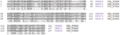
Protein alignment of human hemoglobin proteins, alpha, beta, and delta subunits respectively. The alignments were created using UniProt's alignment tool available online.
-
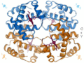
A schematic visual model of oxygen-binding process, showing all four monomers and hemes, and protein chains only as diagrammatic coils, to facilitate visualization into the molecule. Oxygen is not shown in this model, but, for each of the iron atoms, it binds to the iron (red sphere) in the flat heme. For example, in the upper-left of the four hemes shown, oxygen binds at the left of the iron atom shown in the upper-left of diagram. This causes the iron atom to move backward into the heme that holds it (the iron moves upward as it binds oxygen, in this illustration), tugging the histidine residue (modeled as a red pentagon on the right of the iron) closer, as it does. This, in turn, pulls on the protein chain holding the histidine.
-

The giant tube worm Riftia pachyptila showing red hemoglobin-containing plumes
-

Heart of Steel (Hemoglobin) (2005) by Julian Voss-Andreae. The images show the 5-foot (1.50 m) tall sculpture right after installation, after 10 days, and after several months of exposure to the elements.


.jpg)
See also
 In Spanish: Hemoglobina para niños
