Blood smear facts for kids
Quick facts for kids Blood smear |
|
|---|---|
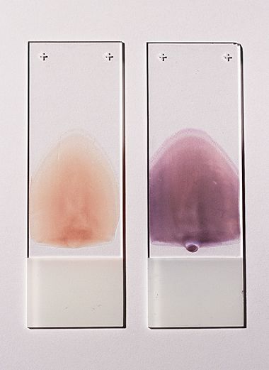
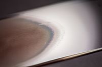
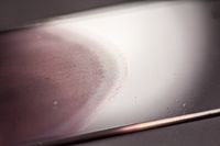
Two push-type peripheral blood smears suitable for characterization of cellular blood elements. Left smear is unstained, right smear is stained with Wright-Giemsa stain.
|
|
| Specialty | {{#statements:P1995}} |
| ICD-9-CM | 90.5 |
| MedlinePlus | 003665 |
A blood smear (also called a peripheral blood smear or blood film) is a tiny drop of blood spread very thinly on a glass microscope slide. It's then colored with special dyes. This lets doctors and scientists look closely at the different blood cells using a microscope. Blood smears help them find out if someone has a blood problem or certain parasites, like those that cause malaria.
Contents
How is a Blood Smear Made?
Spreading the Blood
To make a blood smear, a small drop of blood is placed on one end of a slide. Another slide, called a spreader slide, is used to pull the blood across the first slide. The goal is to create a very thin layer of blood. This thin part is called a "monolayer." In the monolayer, the blood cells are spread out. This makes it easy to count and identify them under the microscope. This perfect area is often found at the "feathered edge" of the smear.
Drying and Staining
After spreading, the slide is left to air dry. Then, the blood is "fixed" to the slide. This means it's briefly dipped in methanol. Fixing helps the cells stick to the slide. It also makes sure the cells look clear when stained.
Next, the slide is stained with special dyes. These dyes make the different types of blood cells show up in different colors. This helps doctors tell them apart.
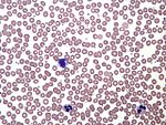
Common stains used are Romanowsky stains. These include Wright's stain, Giemsa stain, or a mix like Wright-Giemsa. These stains help doctors see problems with white blood cells, red blood cells, and platelets. Sometimes, other special stains are used to help diagnose specific blood problems.
Looking Under the Microscope
Once stained, the thin part of the blood smear (the monolayer) is viewed. Doctors use a microscope with very high magnification, up to 1000 times. They look at each cell. They check its shape, size, and how it looks. This information is then recorded.
Why are Blood Smears Important?
Checking Blood Problems
Doctors often use blood smears along with a complete blood count. This helps them check unusual results from blood tests. It also confirms results that a machine might have flagged as unreliable.
Looking at the shape, size, and color of red blood cells is very helpful. It can show what is causing anemia (when your blood doesn't have enough healthy red blood cells). For example, conditions like iron deficiency anemia or sickle cell anemia cause red blood cells to look different on the smear.
Looking at White Blood Cells
The blood smear also helps doctors count the different types of white blood cells. This is called a manual "white blood cell differential." It can show if there are too many or too few of certain white blood cells. For example, it can show neutrophilia (too many neutrophils) or eosinophilia (too many eosinophils).
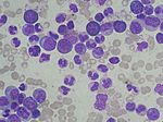
It can also show if there are unusual cells, like "blast cells" seen in acute leukemia. Even modern machines can count white blood cells, but they can't always spot immature or abnormal cells. So, looking at the blood smear by hand is often needed.
Finding Infections
Blood smear examination is the best way to diagnose certain parasitic infections. This includes malaria and babesiosis. In rare cases, doctors can even see bacteria on the blood smear if a patient has a very serious infection called sepsis.
Blood Smears and Malaria
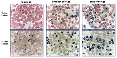
Looking at blood smears is the most reliable way to diagnose malaria. This is because each of the four main types of malaria parasites looks different. Two types of blood smears are usually used:
- Thin smears are like regular blood films. They help identify the exact type of malaria parasite. This is because the parasite's shape is kept very well.
- Thick smears let the doctor look at a larger amount of blood. They are much more sensitive, about eleven times more than thin films. This makes it easier to find low levels of infection. However, the parasites can look more distorted, making it harder to tell the species apart.
An experienced person can find all parasites on a thick smear. But it can be tricky to tell the species apart. This is because the early forms of all four species look very similar. You can't usually identify the species from just one early parasite.
Important Tips for Malaria Smears
It's very important to make the blood smear soon after taking the blood sample. If the blood sits for too long, the malaria parasites can change how they look. For example, if the blood cools down, some parasites might release tiny moving parts. These can be mistaken for other germs.
Also, if the blood sample is left for several hours, especially with certain anticoagulants (chemicals that stop blood from clotting), the parasites can shrink or change shape. This can make them look like a different type of malaria parasite. The best anticoagulant to use is EDTA.
The stains used for malaria blood films also need a specific pH (a measure of how acidic or basic something is). Malaria blood films must be stained at pH 7.2. If the pH is wrong, certain important features of the parasites won't be seen.
If a lab doesn't have experts in looking at blood smears, they can use other tests. These are called Immunochromatographic capture procedures or rapid diagnostic tests. They can quickly detect malaria without a microscope.
See also
 In Spanish: Frotis de sangre para niños
 | Chris Smalls |
 | Fred Hampton |
 | Ralph Abernathy |


