Eye facts for kids
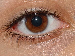
The eye is a special organ that helps animals see the world around them. It works by sensing light. About 97 out of every 100 animals have eyes! Animals like jellyfish, snails, fish, worms, and insects all have eyes that can form images.
In mammals, like humans, the eye has two main types of cells that sense light: rods and cones. These cells send signals through a special cable called the optic nerve to the brain, which then creates the picture we see.
Some animals can see types of light that humans can't. For example, some can see ultraviolet light (like bees) or infrared light (like snakes).
The lens at the front of your eye works a bit like a camera lens. Tiny muscles inside the eye can change its shape, making it flatter or rounder. This helps your eye focus on things that are far away or very close. As people get older, their eyes might not focus as well. Many people also need eyeglasses or contact lenses to help them see clearly.
Contents
Different Kinds of Eyes
There are many different ways eyes have developed to help animals see. Most ways of seeing have appeared more than once in nature's history.
One way to sort eyes is by how many "chambers" they have. Simple eyes have just one chamber, sometimes with a lens. Compound eyes are made of many tiny chambers, each with its own lens, all packed together on a curved surface.
Simple Eyes
Simple eyes are usually made of one light-sensing part.
Pit Eyes
Pit eyes are like small dents or cups in an animal's skin. This shape helps the animal figure out where light is coming from. It's a very basic way to see direction.
These simple eyes are found in many different animal groups. They were probably the first type of eye to evolve. Pit eyes are very small, often with only about a hundred cells. The deeper the pit and the smaller the opening, the better the animal can tell where the light is coming from.
Pinhole Eye
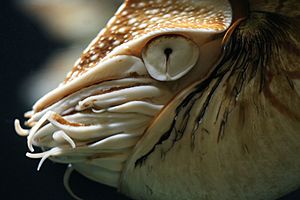
The pinhole eye is a more advanced type of pit eye. It has a very small opening and a deep pit. The Nautilus, a sea creature, is the only animal known to have this type of eye. Because it doesn't have a lens, the image it sees is a bit blurry. The Nautilus can't tell objects apart if they are too close together. A smaller opening would make the image sharper, but it would also let in less light.
Lensed Eye
Adding a lens to a pit eye makes vision much clearer. The lens helps to focus the light, making the image sharper. Some snails and worms have these basic lensed eyes. More advanced lensed eyes have lenses that are thicker in the middle and thinner at the edges, which helps focus light even better.
Many animals with lensed eyes also have special muscles that help keep their eyes steady. This stops the image from blurring when the animal moves.
Some insects have simple lensed eyes called ocelli. These eyes don't form a super sharp image. Instead, they are very good at noticing quick changes in light. This fast response helps insects like flies know if they are flying upside down, as light (especially UV light) usually comes from above.
Reflector Eyes
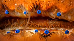
Instead of a lens, some eyes use tiny mirrors inside them. These mirrors reflect light to a central point, creating an image. If you look into such an eye, you might even see your own reflection!
Many tiny creatures, like rotifers and flatworms, use this design. Some larger animals, like scallops, also have reflector eyes. A scallop can have more than 100 tiny mirror eyes along the edge of its shell. These eyes help it spot moving objects.
Compound Eyes
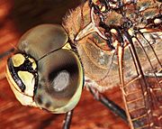
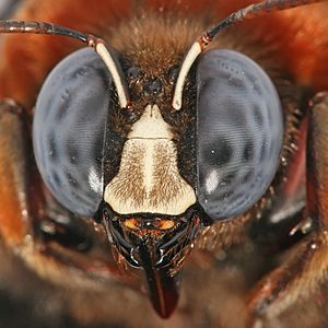
Compound eyes are very different from simple eyes. Instead of one main light-sensing part, they are made up of many tiny units. Some compound eyes have thousands of these units! Each tiny unit is called an ommatidium (many are called ommatidia). The animal's brain puts together the signals from all these ommatidia to create a full picture.
Since each ommatidium points in a slightly different direction, compound eyes have a very wide view. They are also excellent at detecting fast movement and can sometimes even see the polarization of light.
Compound eyes are common in insects, spiders, and some molluscs. Good flyers like dragonflies and praying mantises have special areas in their compound eyes that give them very sharp vision.
How Eyes Evolved
The evolution of eyes started with very simple light-sensitive spots in tiny, single-celled organisms. These early "eye-spots" could only tell if it was light or dark. Many animals use these simple spots to set their internal daily clock, called the circadian rhythm. For example, some snails don't see images, but they sense light, which helps them stay out of bright sunlight.
Even complex eyes still have this basic function. Special cells in our eyes, called ganglion cells, sense light not for seeing images, but to help our brains adjust our daily rhythms to the 24-hour light/dark cycle of nature. This system even works for some blind people who can't see images at all.
Over time, these simple light-sensing spots became more complex. They developed into cup shapes, which helped animals know where the light was coming from. Eventually, eyes evolved into the round shapes we see today, which can focus light onto the back part of the eye, called the retina, to create a full sense of vision, including color, motion, and texture.
How We See
Sharpness of Vision
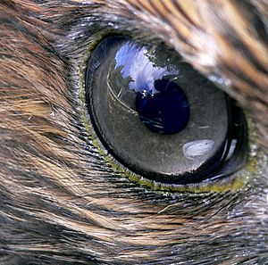
Visual acuity means how well you can see fine details. It's like how clear and sharp a picture looks. For humans, excellent vision means being able to see very small details. A hawk, for example, has much sharper vision than a human, allowing it to spot tiny prey from far away.
Compound eyes, like those of insects, generally have much lower sharpness than the single-lens eyes of mammals. This is because their vision is made up of many small units, which limits how fine the details can be.
Seeing Colors
Color vision is the ability to tell different colors of light apart. Most animals can only see a small part of the light spectrum, mainly between wavelengths of 400 and 700 nanometers. This is a very small section of all the light that exists.
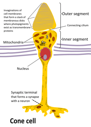
Many animals can't tell colors apart and see the world in shades of gray. To see colors, an animal needs different types of cells that are sensitive to specific ranges of light. In humans and some other animals, these are the cone cells.
Most animals with color vision can also see ultraviolet (UV) light. UV light can be harmful to eye cells. To protect their eyes, many animals have special oil droplets around their cone cells. Humans and some other mammals don't have these oil droplets, so our eye lenses block UV light from reaching the retina.
Rods and Cones
Your retina has two main types of light-sensing cells: rods and cones.
- Rods are amazing at seeing in dim light. They don't see colors, but they help you see in black and white when it's dark. Rods are found all over your retina, but not in the very center.
- Cones are responsible for color vision. They need brighter light to work. Humans have three types of cones, which are sensitive to red, green, and blue light. Your brain mixes the signals from these three types of cones to create all the colors you see. Cones are mostly found in the center of your retina, where your sharpest vision is.
When rods and cones are hit by light, they send electrical signals through other cells in the retina to the optic nerve. The optic nerve then carries these signals to your brain, which turns them into the images you see.
Glasses
Many people wear glasses to help them see better. Glasses are used for vision correction, like for reading or seeing things far away. Sometimes, people wear glasses just for fashion.
Corrective lenses in glasses work by bending the light that enters your eye. This helps to fix common vision problems like nearsightedness (when distant objects are blurry), farsightedness (when close objects are blurry), or astigmatism (when vision is distorted).
As people get older, their eyes often have trouble focusing on close objects. This common condition is called presbyopia. Glasses can help bring the image back into focus on the retina.
Glasses can greatly improve a person's life. They not only make vision clearer but can also reduce problems like headaches or eye strain that come from poor vision.
Images for kids
-
An image of a house fly compound eye surface by using scanning electron microscope
See also
 In Spanish: Ojo para niños
 | Kyle Baker |
 | Joseph Yoakum |
 | Laura Wheeler Waring |
 | Henry Ossawa Tanner |


